-
Ensuring Connection to Better Life!
- Call Us: +91 - 7702199330
- Home
- About Dr. Sai Sudarsan (MBBS, MCh , MS)
- Neurosurgery
- Conditions Treated
- Treatment Offered
- FAQs
- News & Events
- Blogs
- Media Gallery
- Testimonials
- Contact Us
- Our Locations

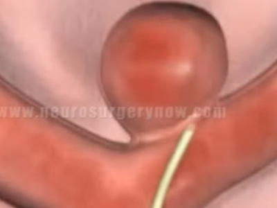
Neurosurgery can be unpredictable and potentially dangerous because of the risk of damage to the nervous system. It is therefore important for surgeons to be able to have someone experienced to monitor nerve tissue during operation. This is called intra operative neurophysiologic monitoring. This monitoring plays a major role in difficult and complex procedures like skull base surgery, curvature correction of spine and cerebrovascular surgery.
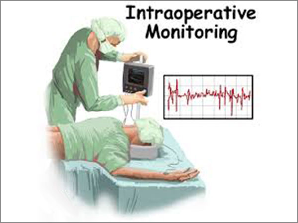
WHAT IS INTRAOPERTIVE MONITORING (IONM)?
IONM uses equipment to evaluate the function of the neural tissue during surgery. Its role is to provide the surgeon with immediate feed back and warning before permanent nerve injury has occurred. It increases the safety and improves the outcome. INOM has been in vogue since mid 1960s. Most neurosurgical procedures bear the risk of permanent neurological injury and din worst cases devastation. In an attempt to reduce such morbidity numerous methods of intra operative monitoring have been created to guide the neurosurgeon in altering operative activity in a way that will prevent or minimize neurological damage.
The ideal intra operative monitoring tool satisfies several technical criteria
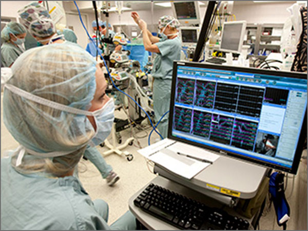
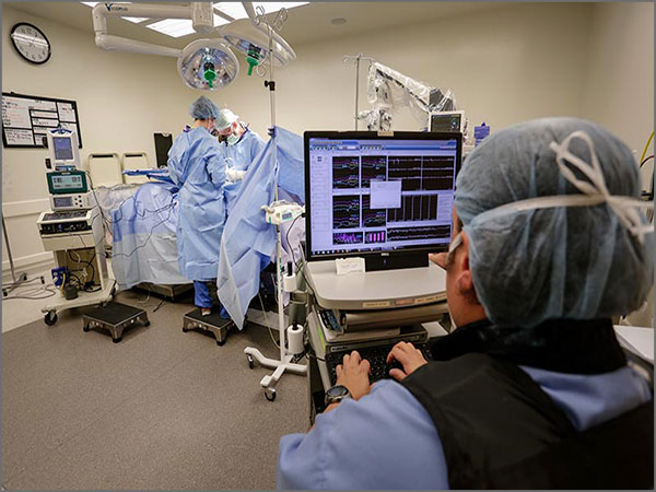
TYPES OF MONITORING
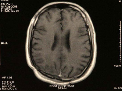
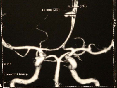
EEG monitoring is being done from 1965. This is a popular mode since it is readily available and is familiar. Other features are simplicity of the pre op set up and well characterized response to various states of neurological function. This modality is commonly used in cerebrovascular surgery like carotid endarterectomy.
Here the recordings are obtained from the brain surface unlike EEG, where recordings are obtained from scalp. ECoG is most widely used in epilepsy surgery to set the boundaries of tissue resection. Chances of seizure free outcome are improved if there is no evidence of seizure activity following resection.
The most widely used technique used in INOM. This measures the conduction of sensation above and below the area of surgery. Electrodes are placed on the limbs that could be affected by surgery and also on the surface of the scalp over the area of brain when the impulses from the limb are received. The activity is recorded as waves. When the limb is stimulated, an electric current there should be a response in the brain.
Here the motor portion is monitored. Needles are placed in the muscle groups that correspond to the area where the surgeon will be working. Electrical activity from the muscle can be monitored by a machine and recorded as waves. Significant change in the waves alerts the surgeon that the neural tissue in the area could be damaged. Surgeon can take action preventing that damage.
Testimonials
Each day is a bonus. It is neither a right nor a natural consequence, unless the all powerful Almighty decides so. Things can go totally haywire in a matter of seconds
MARTIN JOHNSON
Read MoreI am a software engineer based in Hyderabad. I hail from Kerala. I had a cervical disc prolapsed and was advised to undergo surgery at Hyderabad
HARIKUMAR
Read More